

This was an important decision for her because only surgery could offer her the chance of the best possible outcome. After the significance of the results of the NBS mapping session had been explained, she agreed to surgery.
At the start of the operation, invasive direct cortical stimulation of the brain was performed in order to confirm the non-invasive mapping results made with the NBS System. The direct stimulation confirmed the finding from the mapping with the NBS System that the tumor was, indeed, operable. The neurosurgical team was able to remove the tumor, and her clinical outcome exceeded expectations.
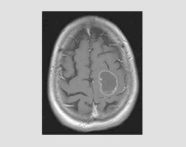
The standard MRI scan showed a tumor in the cortex of the patient's brain. With no information on the location of eloquent cortex responsible for motor function, the treatment decision was difficult.
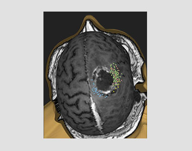
The MRI scan superimposed with results of NBS functional mapping by the NBS System. The NBS mapping results clearly showed that vital motor functions are outside the tumor mass.
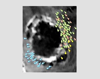
Markers indicate cortical areas where stimuli resulted in responses. Green, yellow and pink indicate areas with responses in hand muscles; blue indicates responses in a leg muscle.
This case illustrates that the Nexstim NBS System can be used for preoperative mapping even in paralyzed (plegic) patients. Even more importantly, this case report demonstrates why tumor resection surgery based on functional NBS mapping information may sometimes lead to substantially better clinical outcomes than surgery planned according to functional MRI (fMRI).
Case report courtesy of Thomas Picht, M.D., Department of Neurosurgery, Charité-Universitätsmedizin Berlin, Germany.