
Nexstim TMS systems with E-field navigation use an algorithm based on mathematically modelling the human brain as over 40,000 spheres. That is over 40,000 individual spheres, rather than one single sphere as in a simple model. Multi-sphere modelling can take into account both the shape and the conductivity of the brain. The unique multi-sphere model used by Nexstim has been scientifically-validated to accurately determine the location and orientation of the maximum induced E-field in the brain.**
If the underlying tissue were homogeneous and perfectly spherical, calculating the position of the induced E-field would be relatively straightforward. However, the shape of the human head and brain is far from spherical and calculating the location requires sophisticated modelling of tissue geometry.
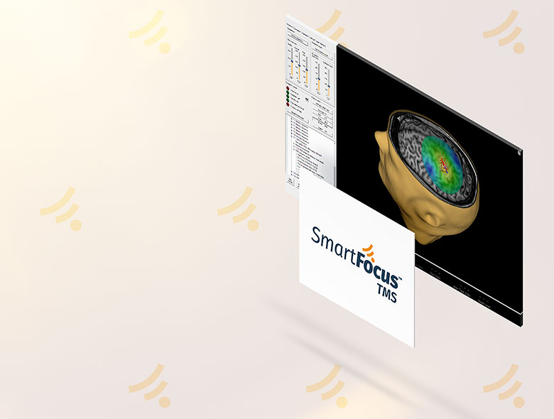
Nexstim system displays the location and orientation of the maximum induced E-field in a 3D rendering, built from the patient’s own MRI head scan by the system's computer. Importantly, the NBS system uses the individual sphere algorithm to dynamically re-calculate the E-field location and orientation. As the operator moves, turns or tilts the coil—even slightly—the system shows the updated location, orientation and E-field data in real time.
Nexstim TMS Systems are the only commercially-available TMS systems with E-field navigation.
TMS, Transcranial Magnetic Stimulation
TMS is a technique for non-invasively transferring an electrical field (E-field) from outside the head into a focussed area of the brain using a pulsatile magnetic field. TMS is normally painless and is rarely associated with serious side-effects*. The magnetic field penetrates the scalp, skull and other tissue without distortion.
Attenuated in the tissues of the head, a TMS pulse can effectively reach at least 3-4 cm below the scalp. This depth allows TMS to be used to probe, or treat, virtually the entire area of the human cortex. And since the cortex is directly and indirectly connected to deeper areas of the brain, stimulation of specific locations can have a profound effect on entire brain systems.
Using figure-of-eight wiring, a TMS coil can be manufactured to have a highly focal effect. In order have a measurable effect, for example depolarize motor neurons, the strength the magnetic field (around 2 T) emitted by a TMS coil need be no stronger than that used in routine MRI scanners.
Navigated TMS
The term navigated has been borrowed from the field of neurosurgery, where the principles of using images of the inside of the brain to guide surgical tools were developed. Originally, this technique was termed stereotactic surgery. Stereotactic meaning "touch in space". With the introduction of frameless stereotactic surgery, where a head tracker also permitted the patient head movement, the term intraoperative neuronavigation became popular among neurosurgeons.
Neuronavigated systems are based on commercially-available tracking systems, which today offer extremely high spatial accuracy. These tracking systems comprise an infrared-based camera, highly-reflective “tracker” spheres and positioning software. The reflecting spheres are fixed on all the tools needed and are also attached to the patient’s head, via a headband, or headframe. Navigation of surgical tools inside the brain is achieved using the data from an MRI scan of the patient’s head. Once a neuronavigated system is “aligned” or co-registered to a patient’s own MRI-scan, the position of any tool can be located, whether it is outside the head or deep inside the brain. An accurate, three-dimensional alignment can be elegantly achieved by matching multiple landmark sites corresponding to the nose, ears and apex of the skull visible on the MRI with the corresponding, real-life landmarks of the patient’s head. Typically, T1-weighted MRI scans with 1 × 1 × 1 mm voxels are used for intraoperative neuronavigation. Requiring less than 1 GB of storage space, these scans are easily portable on various media.
Leaning on decades of experience in neurosurgery, it is a small step to treat a TMS coil as one of the tools to be tracked. This allows for a completely non-invasive neuronavigated system – one which can be used for other applications than intraoperative guidance, for example pre-surgical planning. In principle, coil-navigated TMS only requires adding reflecting spheres to the coil housing, as for any other tool. However, the challenge for tracking TMS is that we are not so interested in the physical location of the coil outside the head, but rather the location of the induced E-field inside the cortex.
The first attempts at neuronavigation of TMS coils assumed that any E-field was induced directly under the center point of the coil windings, a line-of-sight or “line-navigation” approximation. The induced E-field being assumed to be precisely perpendicular to the stimulating magnetic field, which, in turn, is assumed to be precisely perpendicular to the coil winding surface.
Scientific research has, however, shown that coil-navigated TMS using a line-of-sight assumption is a deceptive over-simplification, the human head is far from being a uniform sphere. Coil navigation of TMS has some validity only when the TMS coil is very carefully positioned tangentially against the head before each and every stimulus is given. In practise, coil navigation of TMS can err by up to 10 mm and in worse cases identify the wrong gyrus for the motor cortex.***
Mapping the cortex requires continuous orientation of the TMS coil, including changes in the tilt, as the operator stimulates over the patient's head. Accurately locating the “hand knob” in the motor cortex is important in therapy applications, as setting the stimulation intensity is standardized to the cortical excitability of the patient's primary motor cortex.
Validation of E-field-navigated TMS
The "gold-standard" technique for cortex localization is direct electrocortical stimulation (DES or DCS). DES is a highly invasive procedure. The term “direct” refers to the fact that there is a direct cause-and-effect relationship between the input (E-field) and the output (for example quantitative measurement of muscle EMG amplitude). TMS is the only non-invasive modality that is fully analogous to DES in that both techniques are based on electrical stimulation of a small volume of brain tissue and the direct measurement of the induced effects. The clinical utility of DES is perceived to be very high by neurosurgeons and, although its absolute accuracy is impossible to verify. The accuracy of NBS has been validated by comparing the concordance of pre-operative NBS mapping results with the results of subsequent intraoperative DES in the same patients.
Independent, peer-reviewed articles have shown the ability of NBS to target cortical motor sites to within approximately 2.1 mm compared to DES (Tarapore, 2012). A meta-analysis of all peer-reviewed articles on the accuracy of the NBS System in brain tumor patients found that the mean distance between motor cortex identified by NBS and DES was 6.18 mm (Takahashi et al. 2013), this result is similar in magnitude to the standard deviation intrinsic to DES localization. The FDA cleared the NBS System for mapping the motor cortex in December, 2009, indicating that the device reliably mapped the motor cortex to the same gyrus as the gold standard, DES.
In the 8 years following the FDA clearance of the NBS System, 54 original neurosurgical articles for preprocedural planning covering 2,350 patients were published. These articles included five presurgical motor-mapping studies with a combined total of 633 patients reporting the impact of NBS-derived motor maps on better clinical outcomes**
There is little data on the accuracy of coil-navigated, "line-navigated", TMS. The largest published study comparing line-navigated TMS to E-field-navigated TMS found significantly different results from the two methods, with only a partial overlap of the derived motor maps and, in several patients, line-navigated TMS located the targeted motor hotspot in different gyri compared to NBS.***
The user of an NBS System can be confident that whatever location, angle or tilt of the TMS coil relative to the head, a stimulus with the calculated and displayed E-field will be reliably delivered to the targeted cortical location.
To obtain reliable data and reproducible results, one must be both precise and accurate.
Without accuracy, that is to say without modelling the induced electric field, you can misinterpret the real place of stimulation and stimulate the adjacent gyrus.
Jarmo Laine, MD
VP Medical Affairs, Nexstim
| SmartFocus® TMS – a paradigm shift in TMS therapy for depression |  |  |  |
|---|---|---|---|
| Features | Stand-alone TMS | TMS with coil navigation |
|
| Head/brain image display from MRI | |||
| Coil localization/positioning | / | ||
| Individual modeling of brain conductivity | |||
| E-field orientation shown on cortical anatomy | |||
| Navigation of E-field in cortex | |||
| Integrated motor response (EMG) measurement | |||
| Accuracy clinically proven | |||
| Fully integrated system | |||
| Single & repetitive TMS | |||
FDA-cleared and CE-marked Applications for Nexstim technology |
| ||
| Treatment of major depression disorder (MDD) | |||
| Pre-procedural localization of the primary motor cortex* | |||
| Localization of cortical areas that do not contain essential speech function* |

*Nexstim NBS system with NexSpeech FDA 510(k) K112881
Personalizing the stimulation level for the individual patient
The effects of stimulation are always dependent on the intrinsic state of excitability of the patient’s cortex. Cortical excitability is unique for each individual patient and can vary considerably between patients. When patients have brain disease or stroke, cortical excitability can vary condierably also between the brain's hemispheres.
Once the motor strip has been identified, single-pulseTMS is applied to the hand motor representation area, the “hand knob” with varying intensities to quantify the patient’s resting motor threshold, RMT. A patient’s RMT is defined as the minimum stimulation intensity capable of generating an motor-evoked potential (MEP) in 50% of given stimuli. Nexstim systems use an algorithm to ease the calculation.
The motor cortex needs to be carefully mapped for the optimal hand knob location using all the degrees of freedom available for moving the coil, especially rotation. The goal of this mapping is to locate the site most sensitive to stimulation, otherwise a patient’s resting-state cortical excitability cannot be determined reliably. If the patient’s motor threshold calculation is based on measurement at an erroneous location, there is a risk of over-stimulating the cortex in therapy, as well as causing the patient unnecessary discomfort or pain.
rTMS therapy for depression is normally given at 120% of RMT. Cortical motor mapping can normally be performed at 110% of RMT, or higher.
A TMS primer
References
** Krieg S (Ed.), Navigated Transcranial Magnetic Stimulation in Neurosurgery, Springer International Publishing, 2017
***Sollmann N Comparison between electric-field-navigated and line-navigated TMS for cortical motor mapping in patients with brain tumors. Acta Neurochir (Wien). 2016 Dec;158(12):2277-2289.